You may need to Scroll along >> for full reading.
Red Leg - Caused by Aeromonas hydrophila,
Proteus hydrophilus and Pseudomonas hydrophilus. These types of bacteria are often present in the aquarium but do not affect
the frog. The bacteria is opportunistic and attacks frogs which have a weak immune system or have been stressed. Ulcers and
haemorrhages can often be seen in the legs and belly of the frogs, once these symptoms appear it is usually too late for the
frog. The frogs suffering with red leg often die suddenly without warning. Symptoms of the disease include excess mucus production,
skin discoloration and reddening of the belly and legs of the frog. Treaments that are effective include administering Tetracycline
orally or using Baytril. Adding salt to the water while treatment is going on may increase survival rates of the frogs.
Bloating Disease - common affliction of African Clawed and African Dwarf
clawed frogs. Bloating Disease as it is often referred to is when large amounts of fluids collect in the abdomen, legs and
chin of the frog giving the frog the appearance of a blown up latex rubber glove. The frog can live with this condition
for a short period of time but soon the frog will stop eating and become buoyant due to the pressure of the fluids on the
internal organs. From my research I have discovered that it appears to be caused by the infection of a certain type
of bacteria (still looking into exactly which type) which seems to affect the lymph ducts which drain the bodies fluids properly,
the bacteria seems to block or stop function and ability of these ducts which leads to the accumulation of large amounts of
fluid. This fluid can naturally be broken down by the frogs body if proper conditions or medication is administered.
Aquarium salt and Anti-Internal Bacterial tropical fish remedy has appeared to be successful. Some accounts have
shown that a pure diet of bloodworm can often lead to BD, especially in ADFs. This is perhaps because the bacteria which causes
BD may be present in the digestive tract or epidermis of the bloodworms. To read another article about this by clicking here or here for dwarf frogs bloat.
Tuberculosis - can be caused by organsims such as Mycobacterium xenopi,
Mycobacterium marinum or Mycobacterium ranae. The organisms often gain entry to the frogs body by entering skin wounds or
soars, especially open or bleeding ones. Tuberculosis usually only affects frogs which are already sick from one thing or
another, this is because their immune system is not up to full strength to fight off the disease. Ulcers can occur on the
surface of the skin. Tuberculosis also affects the major organs of the body therefore making the frog very ill. This disease
is not said to be very contagious and is prevented by good hygiene.
Chlamydia psittachi - has symptoms similar to those of bacteria infections
although necrosis of the liver, kidneys, spleen and heart is also present. Tetracycline may be effective in treating this
disease.
Fungal Infections - fungal infections are very common in these frogs
and other amphibians too. Abscesses can be seen in the organs but usually there are ulcers and reddening on the skin, often
with fur like white strands emanating from it. The fungus can be treated successfully with fungicides such as Tetra Fungistop
but internal infections may require treatment with Sulfadiazine. Fungal infection may recur but with proper treatment and
hygiene they can be easily controlled.
Epidermal chytridiomycosis - Afflicting skin. Symptoms include sloughing
and peeling of the skin and extreme buyancy. Malachite gren baths may help.
Parasites -
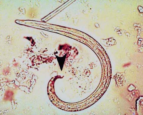
Nematodes such as lungworms (Rhabdias) may cause breathing problems and
pneumonia infections because adult worms live in the frogs lungs, hence the name lungworms (duh!). Egggs and larvae can be
present in the gut. Lungworms can be treated with oral or subcutaneous treatments of 0.2 to 0.4 mg/kg of Ivermectin.
Capillaria infection in the skin will cause skin reddening and irritation,
excess skin shedding, eventually causing death. Salt therapy may be effective.
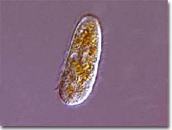
Protozoans - Skin protozoa such as Trichnodina, Costia, Oodinium and Vorticella
often infect ACFs. Symptoms can be skin irritation, cloudiness and excess muus production or skin shedding. Salt therapy may
be effective.
Other Illness -
Gas Bubble Disease is caused when the water in the tank is over saturated
with air. Bubbles can be seen in the foot webbing of effected frogs. Death is not usually caused by this but by the infection
this causes. This is why it is a good idea not to have pumps on for 24 hours a day. Some more info:
The primary problem appears to be gas supersaturation of the water,
meaning
the dissolved gas pressure is greater than atmospheric. This
produces most of the lesions while bacterial invasion
appears to be a
secondary phenomenon.
Experimentally the course of events are:
1. Frogs gradually lose
ability to locate and ingest food
2. Small clear gas bubbles develop within 24-48 h in webbing
3.
Mucus coating on skin reduced
4. Gas bubbles expand in size, number and extent in webbing
5.
Gas bubbles progress to adjacent digits
6. Forelegs preceed hind legs
7. Hyperemia (vascular
congestion) occurs and ascends up legs
8. Hyperemia often accompanied by petechial and ecchymotic
haemorrhages
9. Haemorrhages inclease in size and skin ulcers form
10. Frog unable to stay submerged
and floats to the surface, initially
hind legs up, but after 48-72 h whole frog floats.
11.
Aeromonas can be isolated from some frogs with advanced disease.
Authors were able to reverse signs (if not too severe)
by desaturating the
water.
For static water systems: "water should be allowed to sit for several days
before
animals are added. Gentle aeration can be used to remove gas
supersaturation, but airstones should not be submerged
greater than 0.5 m
Rectal or Cloacal prolapses are where the
linings of the digestive or reproductive tracts extends out of the body, the frogs usually recover spontaneously.